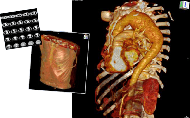
Kenneth Hisley, Ph.D., a
faculty member in DMU
anatomy department, is
manipulating a human torso to
reveal the thoracic cavity of a
human subject – slicing it
horizontally, observing the lungs
and thorax obliquely to show the
spatial relationshipsof the heart
chambers, right and left ventricles,
in relation to a lesion.
Hisley, whose academic interests include clinical imaging
and anatomical visualization, is exploring the thorax not
with physical scalpel and cadaver, but with their digital
equivalents: a sophisticated medical image processing
computer program called OsiriX and an x-ray-computed
tomography (CT) image series of a patient’s thorax and abdomen.
The OsiriX program allows anatomical observation from many viewpoints, zooming in and out of regions, redefining three-dimensional (3D) structural perceptions using tissue colors and transparencies, and the display or removal of arbitrary regions. Hisley sees this new software tool as simply an extension of the physical methods medical students come to know so well, with added advantages for visualizing spatial relationships.
In DMU’s anatomy laboratory, students dissect and explore cadavers at 43 dissection stations, each equipped with a 32-inch flat screen monitor offering dissection directions and descriptive atlas images. Hisley believes this system will be updated to exploit the power of current 3D visualization technology, allowing direct, guided student interaction.
“There are additional manipulations that are difficult or impossible to accomplish with the physical cadaver that might add entirely novel insights into the study of clinical anatomy,” he says.
For example, if a student wishes to understand visceral relationships using an unusual surgical approach, he or she can do so through layers of superficial-to-deep transparencies and without having to turn the physical body. Thus, manipulating CT and magnetic resonance imaging (MRI) image set reconstructions can enhance students’ anatomical knowledge in preparation for their clinical rotations and residencies.
“Physical and digital techniques, by their nature, augment each other,” Hisley says. “Physical dissection provides systematic detailed, spatial and haptic knowledge of anatomy, while digital exploration provides the opportunity for exploration using novel viewpoints increasing recall and a sense of clinical application.”
Previously, Hisley hypothesized that student generation and manipulation of 3D models in parallel with routine physical dissection would significantly improve students’ ability to recognize known anatomy observed from novel viewpoints. He assigned groups of students to explore a randomly assigned region of the body, one group using physical dissection; the other, digital dissection. Results showed that the digital dissectors did better in spatial logic assessment tasks while the physical dissectors were superior in the naming of specific structures. These preliminary outcomes indicate that both techniques should be used in combination.
“Physical dissection is still the gold standard, and I believe it always will be. True anatomical skill is the understanding of complex structures combined with the tactile understanding of the human condition so intrinsic to osteopathic education,” he says. “On the other hand, there is strong evidence that digital anatomical methods are part of the future of clinical practice. Students will need to understand it for this reason as well as for the additional insights it affords into the human body.”
Medical illustration has progressed from ancient times to today, each step evolving in tune with the technology of its day including current 2D, 3D and even 4D images that can be explored and manipulated with digital technology.
Anticipating this need, Hisley is developing an advanced dissector computer program for the dissection laboratory that will enable students to explore their dissection regions using cross-sectional image sequences and 3D models during their practical exercises while affording new methods for real-time student assessment.
Hisley has several additional projects in development in his Biomedical Visualization Laboratory. His advanced auscultation simulator, being developed in collaboration with
the Virtual Reality Applications Center at Iowa State University, synchronizes in real time student
stethoscope placement, location-specific diagnostic sound mixes and 3D anatomic visualization in a series of steps representing the optimal solution to a given diagnostic case, rigorously defined by clinical faculty. Its
goal is to give all students direct access to faculty procedural knowledge patterns regardless of time and distance.

His laboratory student research assistants are working with DMU’s osteopathic manual medicine (OMM) department, chaired by Brad Klock, D.O.’81, to create a 3D visualization model of the thigh’s fascial architecture and dynamics. A better understanding of fascial structural relationships using this digital demonstration model could enrich student understanding of fascial mechanics and OMM procedures.
Hisley’s ultimate goal is to create a DMU-wide online clinical anatomy resource by digitizing and reference-linking a wide range of information resource components (print, image, digital model, sound, etc.) into an indexed library database of training exercises. These accessible learning resources will be intended to execute on all Windows and Macintosh computing tools supporting the curriculum, including desktops, laptops, iPhone/ iTouches and iPads.
A Partnership of federal government and corporations recently launched Text4baby, a new free mobile information service that provides timely health information – in English and Spanish – to pregnant woment and new moms from pregnancy through a baby’s first year.
A Kaiser Permanente study of nearly 35,000 diabetic and hypertensive patients in Southern California, published in the July issue of Health Affairs, found that use of secure patient-physician e-mail communications resulted in a significant improvement in several disease-control measures.


